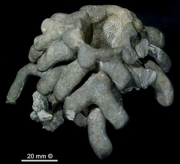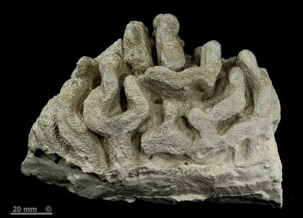
Figure 1 - Aphrocallistes alveolites.
Alemannia, Höver, senonensis zone.
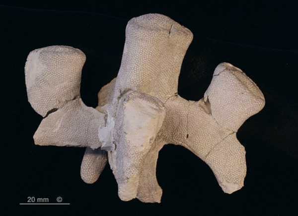
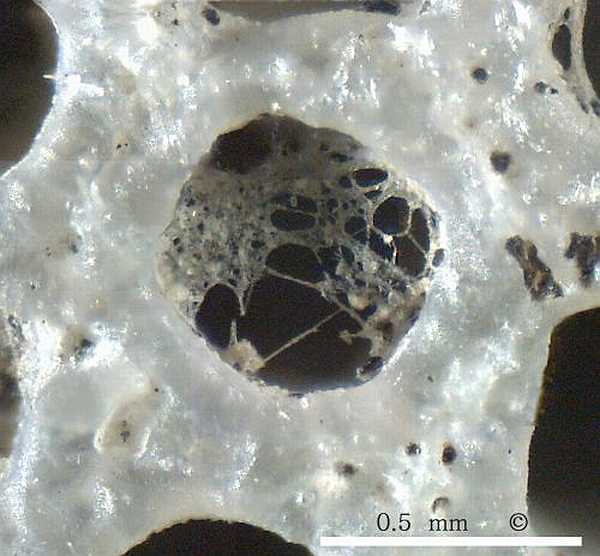
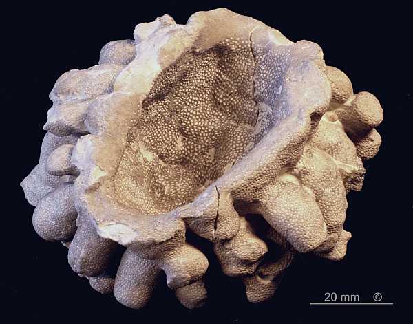
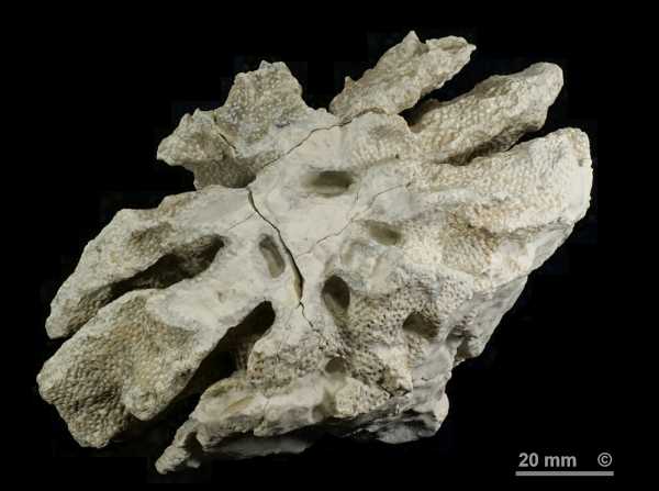
Figure 2 - Aphrocallistes alveolites.
Teutonia North, Misburg, vulgaris/stolleyi zone.
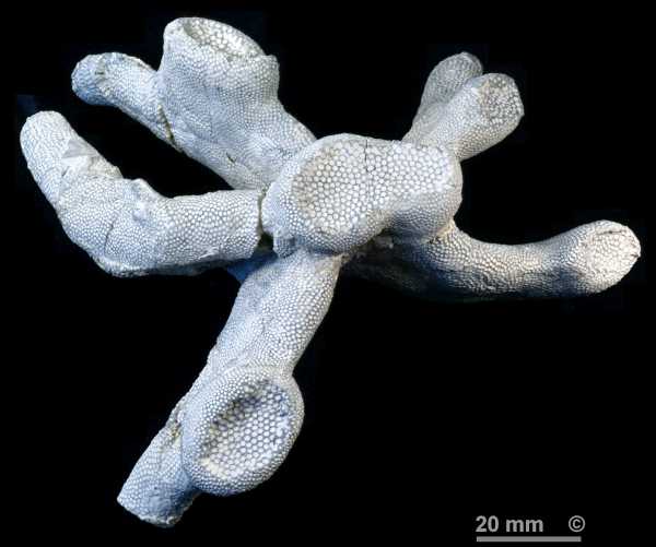
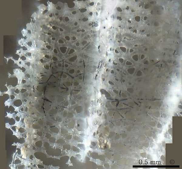
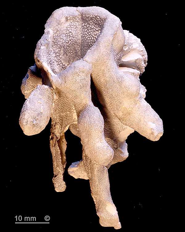
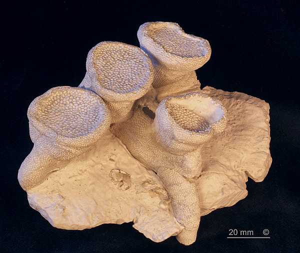
Figure 3 - Aphrocallistes alveolites.
Teutonia South, Misburg, conica/mucronata zone.
Synonyms:
Scyphia alveolites Roemer 1841
Occurence:
Alemannia, Höver, Lower Campanian (senonensis zone). Moderately common.
Teutonia, Misburg, Upper Campanian. Moderately common.
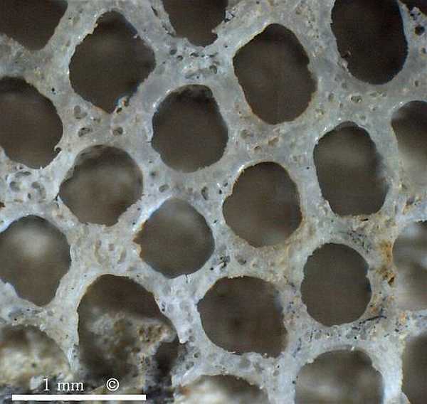
Aphrocallistes alveolites forms subhorizontal branching tube systems, with vertical chimneys whose terminal apertures are covered by sieve plates. The basal tubes are 10 to 20 mm in diameter, the chimneys may be up to 50 mm in diameter. Presently available material suggests that there is generally one central chimney which sends out several radial tubes, each of which has its own secondary chimney.
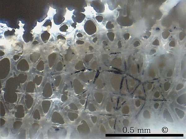
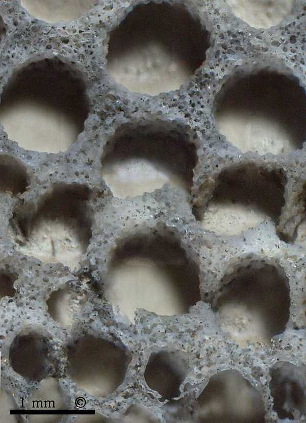
The tube walls are approximately 1.5 mm thick and are perforated by radial channels (diarhyses), giving rise to the very regular, almost honeycomb-like pore structure which makes Aphrocallistes species easily recognizable.
Figures 1, 2 and 3 show fragmental specimens consisting of subhorizontal, branching tubes with closed, finger-like terminations and chimneys. The chimneys have wide oscules covered with characteristic sieve plates whose structure resembles that of the tube walls, but the pore sizes are distinctly larger.
The finger-like ends of the subhorizontal tube sections are rounded, closed, and often point downward and may grade into root-like supports.