Cameroptychium serotinum
Schrammen 1924
Cameroptychium serotinum has been described by Schrammen (1924) from the Lower Campanian of Höver on the basis of a single specimen. Other species of Cameroptychium described by Schrammen (1912) and Leonhard (1897), also based on single specimens only, include Cameroptychium planum from Heere (near Salzgitter, North Germany) and Cameroptychium patella from Opole (Oppeln in older German literature) in Upper Silesia / Poland. Schrammen (1912) also described the new species Camerospongia pervia (based on 2 specimens, no figures given) from Oberg (near Ilsede, North Germany), and his verbal description suggests a great similarity with Cameroptychium serotinum.
Over the years, the author has collected about a dozen samples from Höver, and the accumulated material suggests that these specimens are Cameroptychium serotinum (which may be equivalent to Camerospongia pervia, as which they were regarded before).
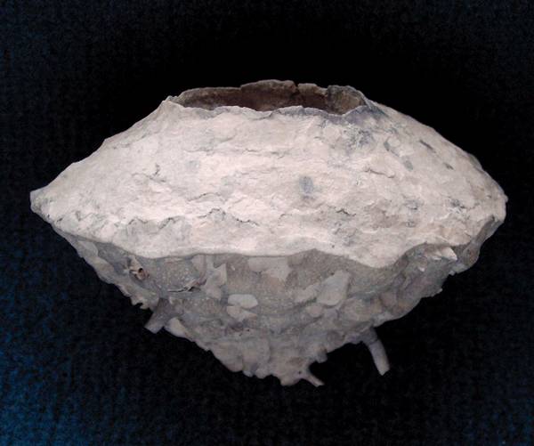
The upper parts of Cameroptychium serotinum usually have the form of a truncated cone, but may show more convex, almost hemispherical shapes in some samples. The siliceous membrane covering the upper part is extremely thin, smooth, and with numerous small pores. The membrane has some gaps which are difficult to discern and which correspond to cavaedial interspaces in the interior of the thick (20 to 30 mm) folded wall.
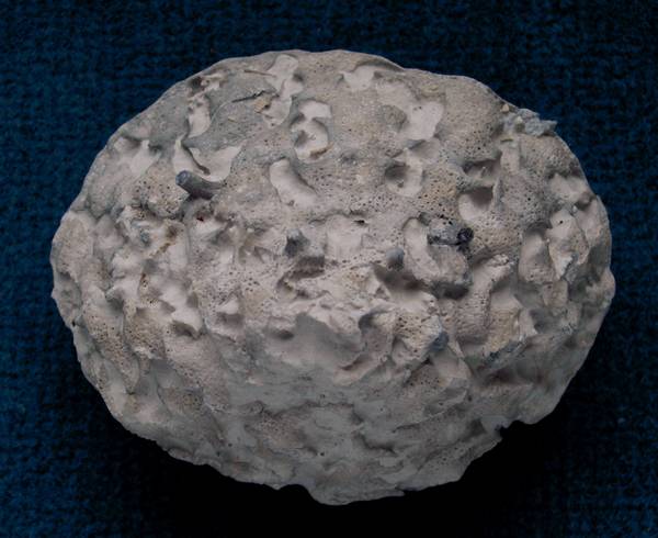
The lower part of Cameroptychium serotinum is obconical and may be almost flat to very acute (more acute than the upper part). The obconical part is stronly sculptured and consists of indistinct, broad (10 mm) and highly irregular, partially interconnected radial bands, separated by irregular cavities. The bands are covered with a strong cortex drived from dermal lychnisks. The cortex has many small (0.3 to 0.5 mm) pores which may show a regular quadratic arrangement locally.
A number of solid, thin (2 to 3 mm) and smooth roots are emerging from the basal cortex (very similar to the roots of Becksia, Discoptycha, and Marshallia).
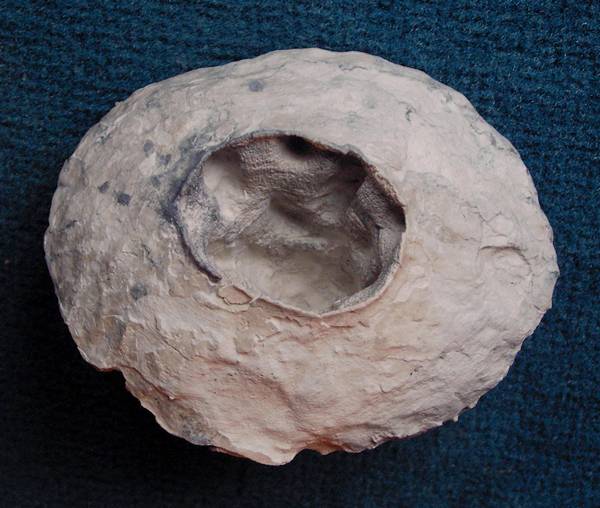
Cameroptychium serotinum has a wide (20 to 40 mm) circular osculum, leading into a deep paragastral cavity. The paragastral surface and the entire wall consist of vertical, upwardly branching (and in some cases reuniting), 5 to 10 mm wide folds.
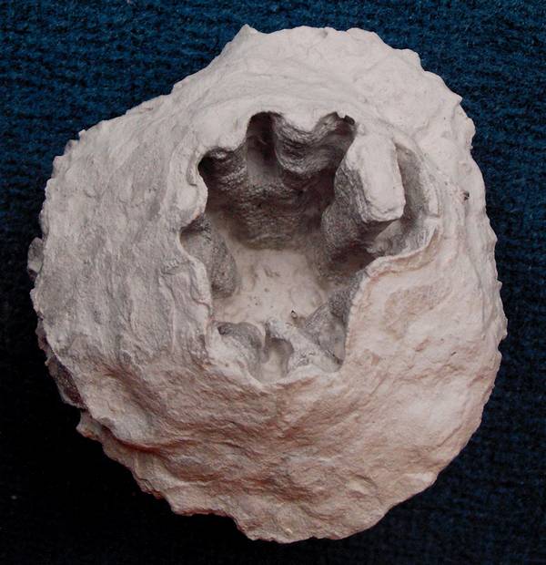
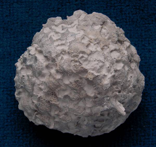
The second example has an osculum with a distinctly sinuous outline and shows a somewhat anomalous, isolated fold which is almost completely closed to form a tube.
Cameroptychium scharnhorsti
Krupp 2010
Cameroptychium scharnhorsti was described as a new species recently by Krupp (2010). The original description may be downloaded here as pdf-file (1.4 MB):
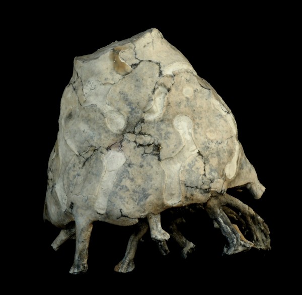
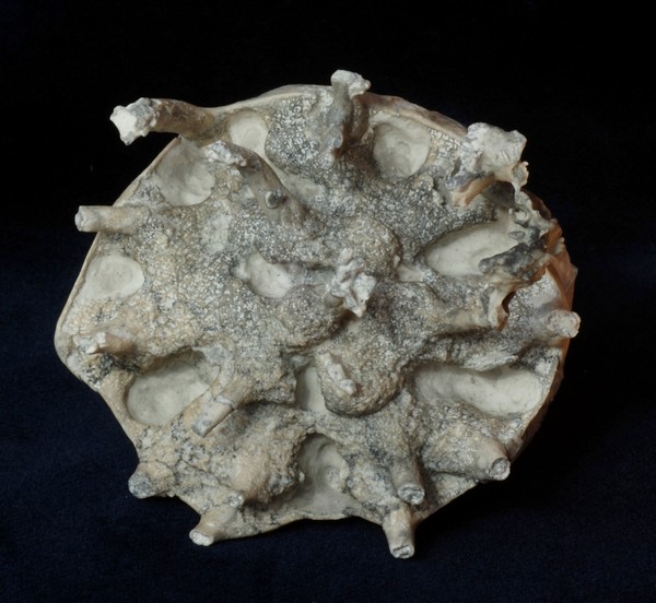
The first and second picture show a complete specimen in side and bottom view. The third picture is another, comparatively small specimen. To date, Cameroptychium scharnhorsti is only known from the Lower Campanian of Höver.
Cameroptychium scharnhorsti has a barrel-shaped to conical habit and a wide (20 to 40 mm) osculum and deep paragastral cavity. The sponge may attain sizes of more than 100 mm in diameter, but typical examples are about half this size.
On the dermal surface, orbicular structures of partially confluent rings are very conspicuous. The rings consist of a finely porous siliceous membrane. These rings encircle the terminations of short tubes which form part of the folded sponge wall. Between these rings and tubes, cavaedial gaps exist in the wall. The base of Cameroptychium sp. is flat to slightly concave and is covered by a rough cortex from which several short root stubs emerge. All these features are in homology to the wall structure of Cameroptychium serotinum, although they differ in detail: The barrel-shaped wall corresponds to the upper conical margin of Cameroptychium serotinum, and the orbicular cortical membrane is more solid than that of Cameroptychium serotinum. The root stubs and the cortical surface of the narrower base of Cameroptychium scharnhorsti are very similar to those of Cameroptychium serotinum.
Also, the tube-forming skeleton of Cameroptychium scharnhorsti is made up of a well ordered lattice of lychnisks with cubic meshes, just as in Cameroptychium serotinum.