Cretaceous sponges from the Campanian of Misburg and Höver
Laocoetis Pomel 1872

Locality: Teutonia. Height: 170 mm

Laocoetis crassipes

Pomel 1872

Laocoetis crassipes has not been reported by Schrammen (1910-1912, 1924) for Misburg and Höver. However, Laocoetis crassipes is probably synonymous with Ocellaria cancellata of Roemer (1864), who described the species from the Cretaceous of North-Germany.

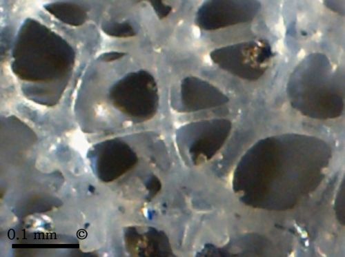
The specimen shown is a very thin-walled (1 mm), slightly folded funnel with a short stalk. The inhalent pores (epirhyses) on the dermal side are approx. 0.5 mm wide and are arranged in longitudinal and transvere rows, forming a rectangular pattern. The pattern of aporhyses on the gastral side is very similar, but the aporhyses are offset in such a manner that they occupy the positions between four adjacent epirhyses (arranged in quincunx).

Locality: Teutonia. Height: 170 mm
Locality: Teutonia. Height: 240 mm

Laocoetis sp.

new species

Laocoetis sp. is probably a new species from Misburg, known only from the Upper Campanian so far.

The new sponge forms chimney-like, thin walled (2-5 mm), cylindrical tubes, typically 30 to 50 mm wide. Typically, chimneys are side branches debouching from a common, similar sized, horizontal tube at their base.

The tubes have a conspicuous regular pore pattern with pores arranged strictly in longitudinal rows, and less strictly in transverse rows. Typical pore diameters are 0.5 mm, and pores are separated from their neighbors by about the same distance. However, pore size appears to vary considerably between individual specimens. The pore pattern of the gastral side is very similar to that of the dermal side.

Both gastral and dermal surfaces show a cortical meshwork formed from dictyonal hexactines mantled by additional silica. Upon acid treatment, the reinforced dermal and gastral layers tend to separate along the weak internal skeletal layer.

Photomicrograph, dermal pores

Photomicrographs of Laocoetis sp.

The first image shows the typical rectangular arrangement of inhalent pores on the dermal side of the sponge. The dermal hexactines are mantled by additional silica, forming a rigid, superficial meshwork.


The second image is an inside view of the dermal side, showing longitudinal strands of strictly aligned, dictyonal hexactines between longitudinal rows of epirhyses.

The third image shows a cross section (perpendicular to tube axis; dermal side on the left), showing several epirhyses.

Photomicrograph, dermal pores

Photomicrograph of Laocoetis sp.

Framework of dictyonal hexactines between epirhyses. Some hexactines have arms reduced to acute cones (middle left marging).


Photomicrograph, hexactines

Photomicrographs of Laocoetis sp.

The first image shows a section of the dictynal skeleton of Laocoetis sp., composed of small hexactines with smooth surfaces.

Occasionally, dictyonal hexactines have appendices made up of smaller hexactines like the ones in the two next images.


BACK
HOME