Leiochonia cryptoporosa
Schrammen 1901
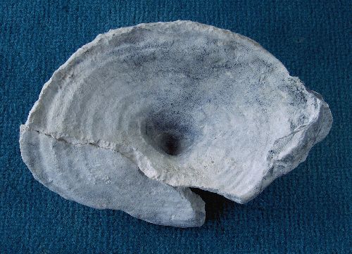
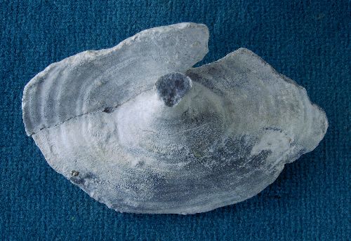
Leiochonia cryptoporosa is a very rare species at Höver. It occurs as flat funnel- to ear-shaped individuals. It is distinguished from the slightly more common Leiochonia robusta by its minor wall thickness, but this distinction seems somewhat arbitrary.
The specimen shown here from above and below has been broken, probably during early diagenetic compaction of the sediment. The wall thickness is 6 to 8 mm, and both sides show distinct concentric growth stage marks. The rim is straight, with rectangular edges against the gastral and dermal surfaces.
Leiochonia robusta
Schrammen 1910
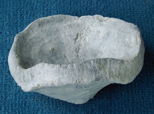
Leiochonia robusta is a rare species at Höver. It occurs as flat funnel- to ear-shaped individuals. The funnels often show a slightly undulating rim and a tear-shaped outline with a v-shaped notch.
The specimen in the picture shows the typical broad margin, forming distinct edges against the dermal and gastral surface. The margin also shows some typical anastomed furrows.
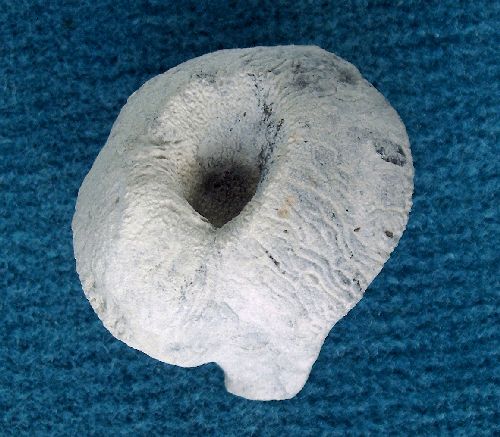
The second example of Leiochonia robusta shows nicely the characteristic over-proportional wall thickness and the anastomosing furrows on the margin. Notice also exposed pore channels inside the funnel. More often the gastral and dermal surfaces of Leiochina are completely smooth, a fact that gave rise to the species name cryptoporosa.
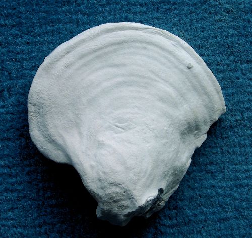
Dermal side of an ear-shaped specimen of Leiochonia robusta, showing typical concentric growth rings and the common smooth development of the surface without noticable pores.
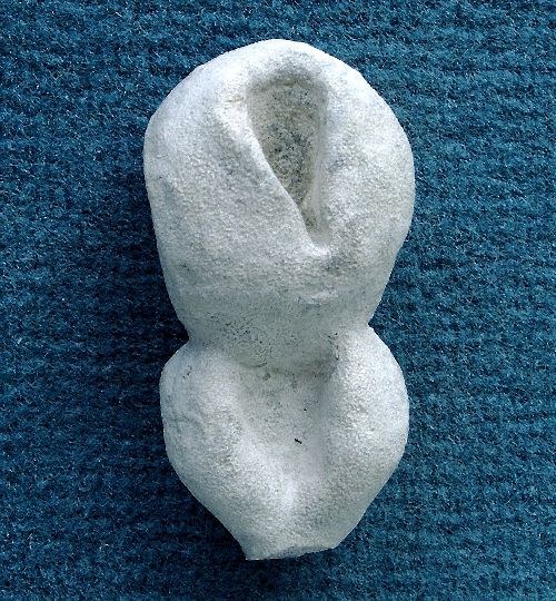
A small specimen of Leiochonia robusta, showing typical tear-shape and reproduction by budding.
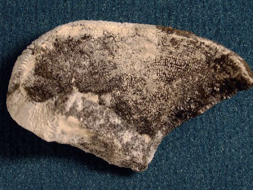
Etched specimen of Leiochonia robusta, revealing internal canalization. Notice that the aporhyses occur in layers corresponding to concentric growth zones.
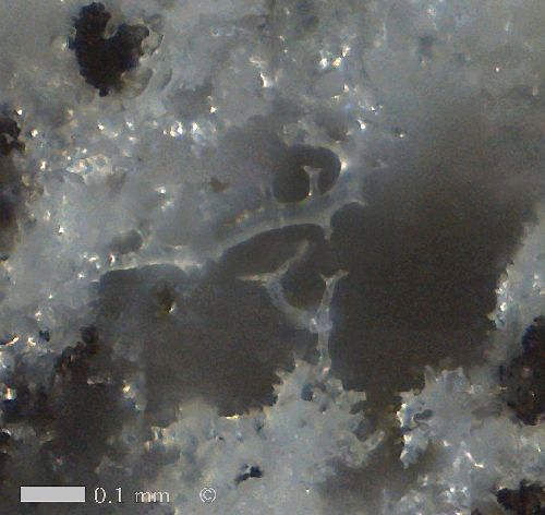
Epirhysis of Leiochonia robusta, traversing a dense skeleton of rhizoclones. Inside the canal, two rhizoclonids.
Rhizoclonids are only observed inside of canals, suggesting that they may have functioned as supports for choanocyte cells.
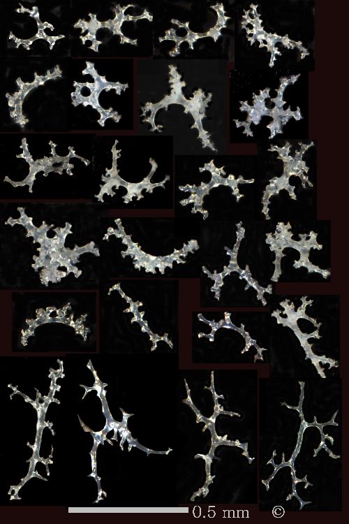
Leiochonia robusta. Selection of typical rhizoclones (rows 1-5), and rhizoclonids (row 6).
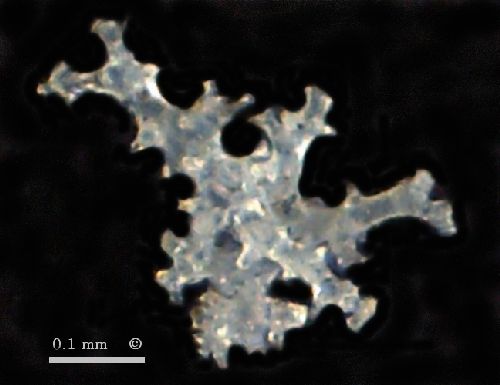
Compared to other rhizomorina, the skeleton-forming rhizoclones of Leiochonia are very small (0.2 to 0.3 mm). The rhizoclones are sturdy, with curved, straight, or branching morphologies and with many short clones (root-like appendices).
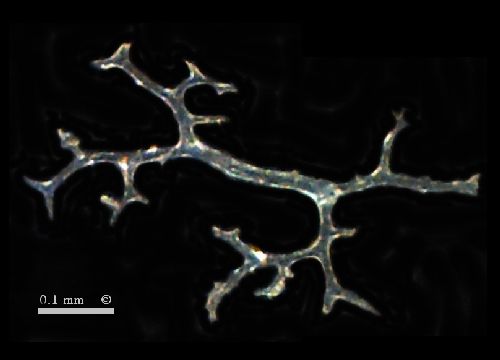
Besides the rhizoclones, there are also much less abundant rhizoclonids. The latter are distictly larger than the rhizoclones, have a stretched appearance with smooth surfaces and with few, well developped arms, often with bifurcated terminations.
The next two images show details of a typical rhizoclone and a typical rhizoclonid.