
Lepidospongia fragilis
Schrammen 1902
Lepidospongia fragilis is a moderately common hexactinellid sponge in the Upper and Lower Campanian of the Misburg area, but is hardly ever found as complete specimens.
Typically, Lepidospongia fragilis forms regular funnels with a stem, or more irregular shapes. From other specimens it is known that the species has strong, branching roots.
The name Lepidospongia alludes to scaly fish skin, describing the tiled appearance of the gastral surface. Under the microscope, the "scales" (siliceous plates formed from modified dermal lychnisks) reveal a typical perforated pattern (see below).

Specimen of Lepidospongia ?fragilis, showing typical dermal surface with deeply entrenched and narrow interspaces between parallel folds. (Stem of sponge occupied by a brachiopode)
Similar dermal surface strctures are observed with several other hexactinellid sponges, making identification difficult unless the gastral surface of the specimen can be exposed.

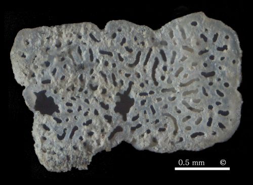
Enlarged view of the first specimen (top image), showing gastral surface with siliceous platelets.
These delicate perforated plates are the distinguishing feature of Lepidospongia fragilis.

Perforated siliceous platelets from the gastral surface of Lepidospongia fragilis. (Photomicrographs of scleres isolated by dissolving a sample in acetic acid.)

Lepidospongia rugosa
Schlüter 1870
Lepidospongia rugosa appears to be more common than Lepidospongia fragilis, and it tends to form larger individuals. Complete specimens are unknown to the author.
Lepidospongia rugosa occurs as regular (small) trumpet-shaped funnels to fairly large and irregular, flat funnels. The species shows a similar gastral covering layer as Lepidospongia fragilis, but the tiled appearance of the latter is lacking. Instead, the gastral surface of Lepidospongia rugosa has a rough appearance, with some concentric banding.

The example of Lepidospongia rugosa shown here is a fragment of a large flat funnel. It shows several conical to finger-shaped prominences emerging from the gastral surface, or near the funnel margin.
Occasionally, such isolated conical or finger-like fragments of Lepidospongia rugosa and Lepidospongia fragilis are found, that show the typical gastral surface features on their outsides (!), which may be confusing.

Etched sample of Lepidospongia rugosa showing folded dermal surface with narrow trench-like interspaces.
The ostia are aligned along the bottom of these trenches. They are generally covered by sediment and therefore invisible. On the trench walls numerous spiny prominences emerge from dermal lychnisks and provide a sieve mechanism to prevent clogging of the inhalent canal system.

Photomicrograph of etched sample of Lepidospongia rugosa showing folded dermal surface with narrow trench-like interspaces. On the bottom of the trenches the ostia are recognizable (slightly out of focus).
The lychnisks forming the fold ridges are mantled and reinforced by siliceous material which also reduces the mesh apertures to tiny round pinholes.

Photomicrograph of etched sample of Lepidospongia rugosa showing siliceous membrane covering the gastral surface.
The silicous membrane is more or less continuous (no tiles, as with Lepidospongia fragilis), although it is rarely preserved in an undamaged state. The postica are formed by oblique openings reminiscent of eye lids.

Detailed view of a lid covering a postica. The siliceous membrane has a faintly stippled ornamentation.

Internal view of a fold, showing fairly regular cubic grid of lychnisk scleres.
In the bottom left part, the siliceous covering layer of the dermal surface can be recognized behind the lychnisk grid.
Along the top margin, the image shows the outside of the dermal surface. Notice the spiny protrusions formed by the outward pointing, mantled lychnisks arms.

Detail of the skeleton made up of lychnisk scleres.
Notice the appendices on unmated lychnisk arms, forming the walls of aporhyses.