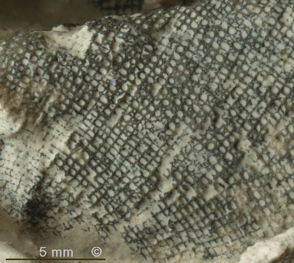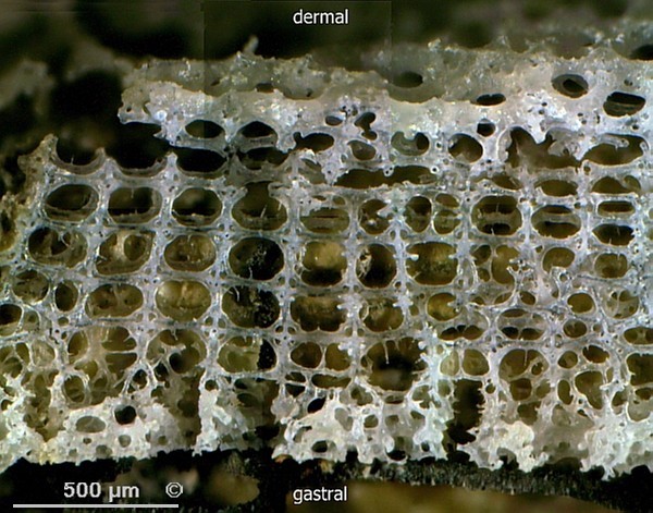
Figure 1 - Marshallia tortuosa, Alemannia, Höver.
Basal section and stem.
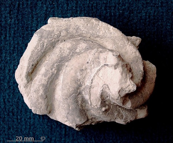
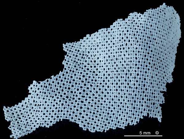
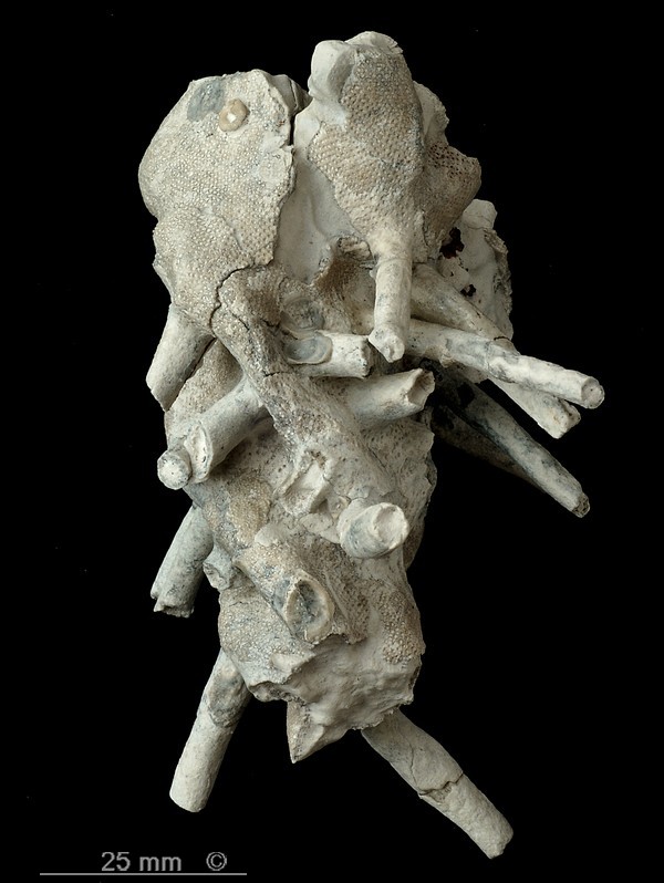
Figure 2 - Marshallia tortuosa, Alemannia, Höver.
Mid section, viewed from below.

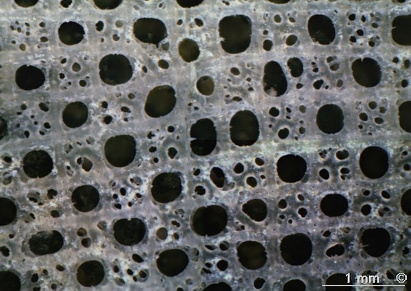
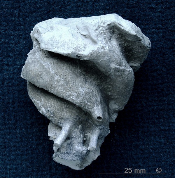
Figure 3 - Marshallia tortuosa, Alemannia, Höver.
Stem and root processes.
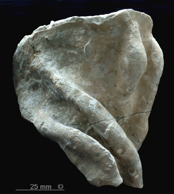
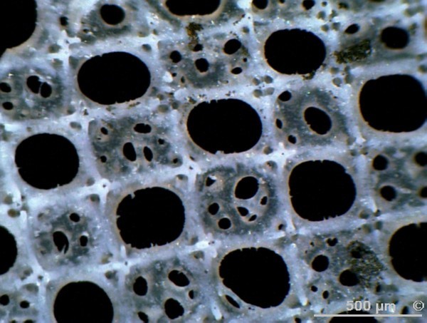
Figure 4 - Marshallia tortuosa, Alemannia, Höver.
Top section with smooth brim.
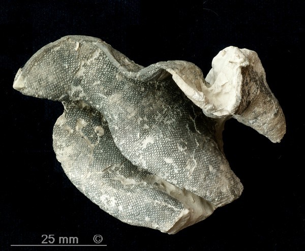
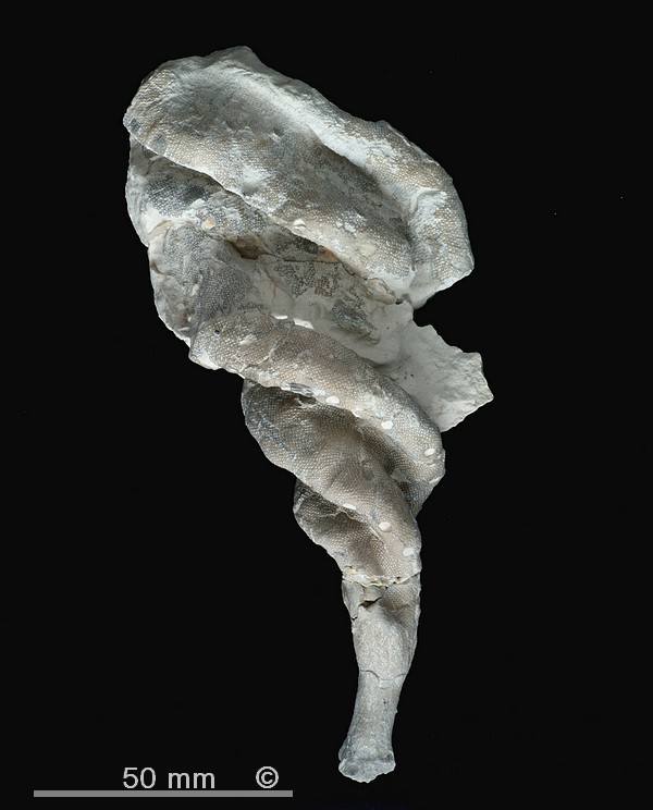
Figure 5 - Marshallia tortuosa, Alemannia, Höver.
Top section with a sinuous margin.
Synonyms:
Pleurostoma tortuosa Roemer 1864
Spirolophia tortuosa Pomel 1872
Occurence:
Alemannia, Höver, Lower Campanian (senonensis zone). Rare.
Marshallia tortuosa is a moderately rare species of the Lower Campanian of Höver. Apparently, a second Marshallia species exists, which occurs in both the Lower Campanian of Höver and the Upper Campanian of Misburg. It will be described informally below.
Marshallia tortuosa has a very characteristic twisted habit (Figures 1, 2, 4 and 5), resulting from a spiralling arrangement of longitudinal (actually oblique) folds in the funnel wall. The individuals can look quite different, depending on the number of folds, which can range from two to at least five. The specimen shown in Figure 1 has three folds and a slender habit, while that in Figure 2 has five folds and a flat shape.
Marshallia tortuosa has a proper stem with laterally radiating roots at its base (Figures 1 and 3).
In its lower sections, the fold crests of Marshallia tortuosa show accessory oscula, which are circular and 3 to 4 mm in diameter (Figures 1 and 4). Near the upper end of the sponge, intercalated secondary folds may develop (Figure 4).
The upper brim of Marshallia tortuosa may be smooth as in Figure 4, or sinuous as in Figure 5.