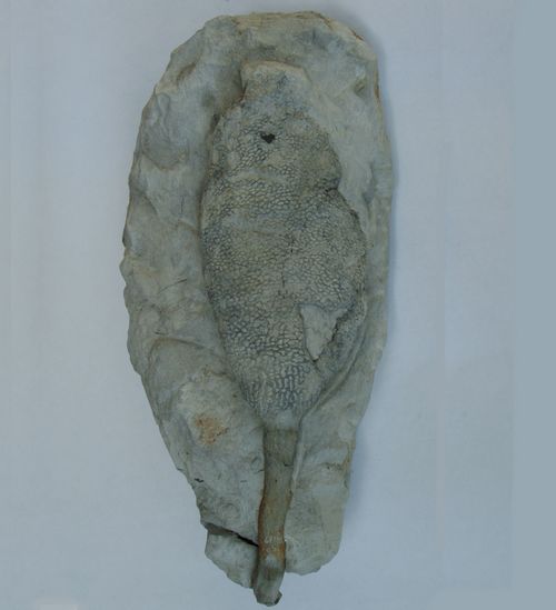
Sporadoscinia micrommata
Roemer, 1841
Sporadoscinia micrommata is a vase-shaped, fairly thin-walled (2 mm) sponge. Its stem is quite long and the transition into the cup is rather abrupt. The outside surface shows a characteristic irregular network of more or less polygonal pores whose diameteres are smaller in longitudinal than in circumferencial direction.
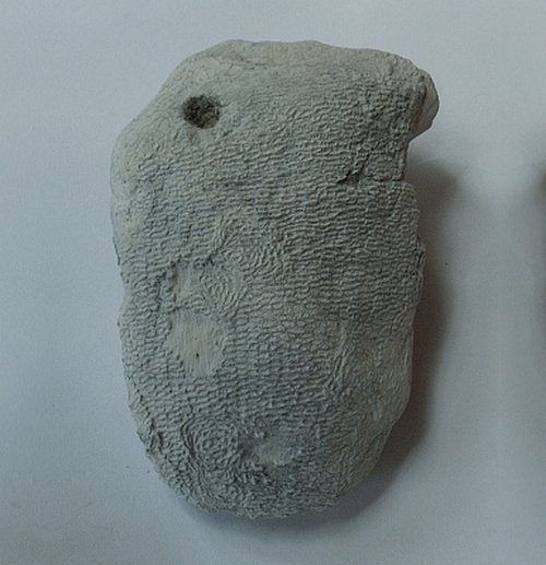
Isolated cup of Sporadoscinia micrommata, with typical circular spots in the otherwise subparallel mesh fabric of the wall. These spots are thought to be repaired holes, probably inflicted by a predatory organism. There is also a similar sized, unrepaired hole near the left top margin.

Fragment of Sporadoscinia micrommata, viewed from outside (left) and inside (right). Notice the regular, longitudinal, in quincunx arrangement of oval postica versus the highly irregular and angular ostia.
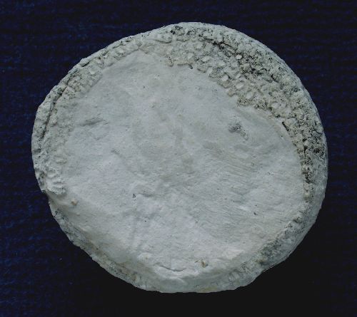
Top-view of a specimen of Sporadoscinia micrommata, showing some relics of what appears to be a sieve plate, covering the funnel aperture. Sieve plates have not been observed before with Sporadoscinia species.
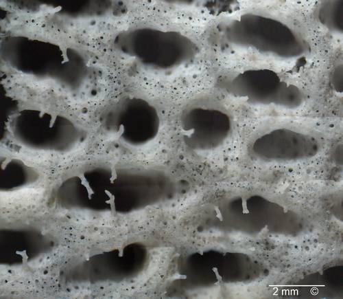
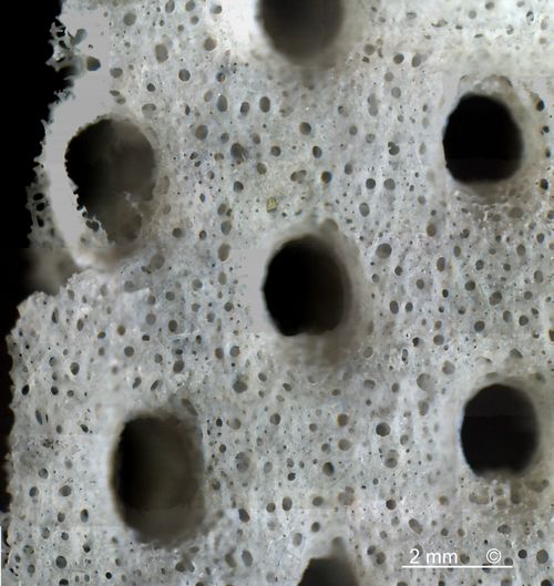
Etched fragment of Sporadoscinia micrommata, viewed from outside (top) and inside (bottom).
Notice finely porous membrane formed by dermal and gastral spicules between the pores. Also notice the root-like skeletal processes in front of the ostia (upper image), which probably served to prevent clogging of pores by larger particles.
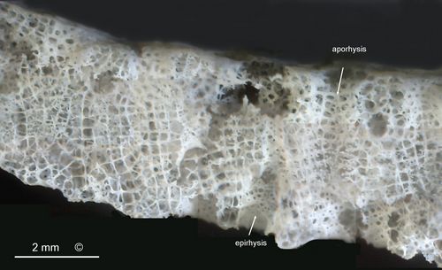
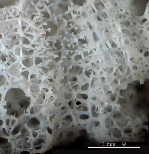
Top: Etched cross-fractured specimen of Sporadoscinia micrommata, showing internal wall structure. The image shows the dictyonal skeleton consisting of lychnisk spicules, which enclose alternating inhalent and exhalent passages (epirhyses and aporhyses) in the sponge wall. The epirhyses are lined with a siliceous porous membrane while the aporhyses consist of open passages through the dictyonal skeleton.
Bottom: As above, but at higher magnification. View of the interior of a vertical aporhysis in the center, and onto the exterior of two epirhyses near the left and right margins. The epirhyses have walls consisting of a porous siliceous membrane, while the aporhyses have no real walls, but are lined with root-like endings of dictyonal lychnisks.
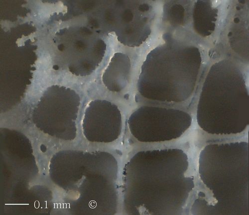
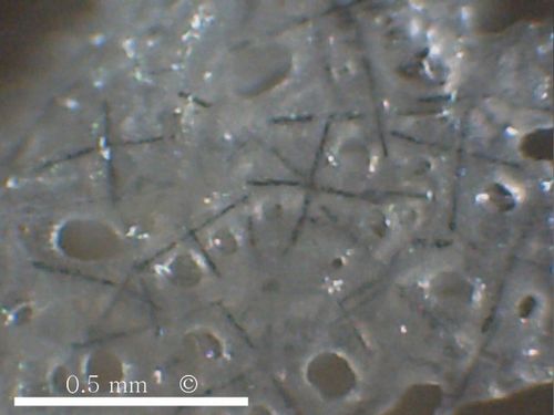
Top: dictyonal skeleton of Sporadoscinia micrommata, consisting of smooth to barbed, fused lychnisks.
Bottom: Detail of siliceous membrane (gastral side), showing black axial canals of gastrals lychnisks. Notice how the siliceous membrane spans between the lychnisk arms.

Sporadoscinia quenstedti
Schrammen 1912
Sporadoscinia quenstedti is a rare sponge at Misburg. The almost completely preserved specimen from the Teutonia pit shown in the picture is bedded on a large fragment of Ventriculites radiatus.
Due to the very meagre description in the literature, the proper identification as Sporadoscinia quenstedti (and not as Cinlidella solitaria, as originally assumed) was only possible after comparison with the type specimens preserved in the collection of the Institute of Paleontology in Göttingen.
The pores of Sporadoscinia quenstedti have a tendency to arrange in a rectangular pattern. Where the ridges between the pores intersect, a pronounced knob is formed.
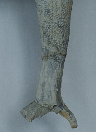
The transition from the poriferous body of Sporadoscinia quenstedti to the short stem and roots (lower 70 mm) is rather sharp (within 2 millimeters). The root is composed of densely aligned fibres, apparently derived from distorted skeletal lychnisks (bottom picture).
Sporadoscinia teutoniae
Schrammen, 1912
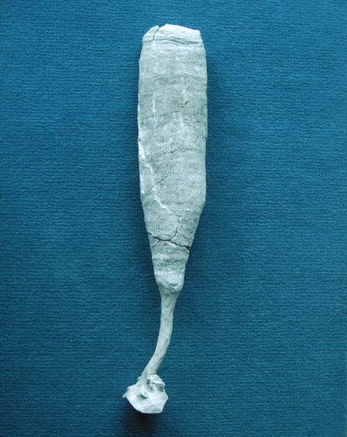
Sporadoscinia teutoniae has a tube-like habit, tapering downward, but the uppermost rim is often slightly constricted. The transition from the porous tube into the stem is morphologically smooth and continuous, not stepped. The stem is fibrous. The dermal surface of the tube-section shows an irregular network of more or less polygonal pores, but the pores tend to be smaller (1 mm) than those of Sporadoscinia micrommata (1.5 to 2 mm), and the bridges between the pores are plane. Sporadoscinia teutoniae is similar to Leiostracosia sp., but the latter has its postica arranged in longitudinal rows.

...Sporadoscinia teutoniae
Sporadoscinia venosa
F.A. Roemer 1841
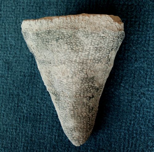
Sporadoscinia venosa is distinguished from the similar S.micrommata by its smaller ostia, and by wider bridges between them. Also the bridges are flat and not protruding. The ostia tend to be arranged in longitudinal rows.

This virtually complete specimen of Sporadoscinia venosa was recovered in June, 2007. It shows a constricted growth margin at the top and a thin short stem with laterally branching roots.

Another fine specimen of Sporadoscinia venosa.
Sporadoscinia decheni
Goldfuss 1826
Sporadoscinia decheni is much rarer than the other Sporadoscinia species. Stratigraphically, its occurrence is limited to the Lower Campanian.
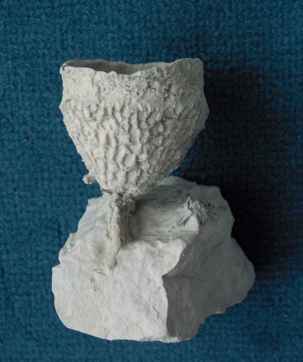
The specimen shows a small but excellent example of Sporadoscinia decheni. The species is distinguished by its large, angular, irregularly arranged ostia and the angular bridges in between. Typically, it occurs in shallow, cup-like forms. The specimen shown here has only a very short stem.
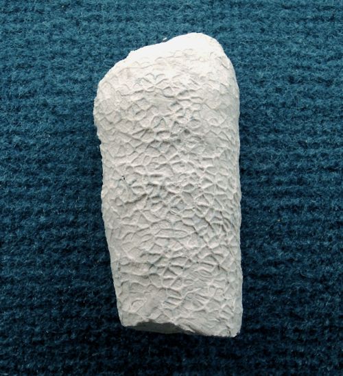
Besides the typical cup-shaped specimens of Sporadoscinia decheni, a more tube-shaped variety also occurs.