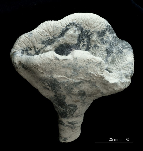
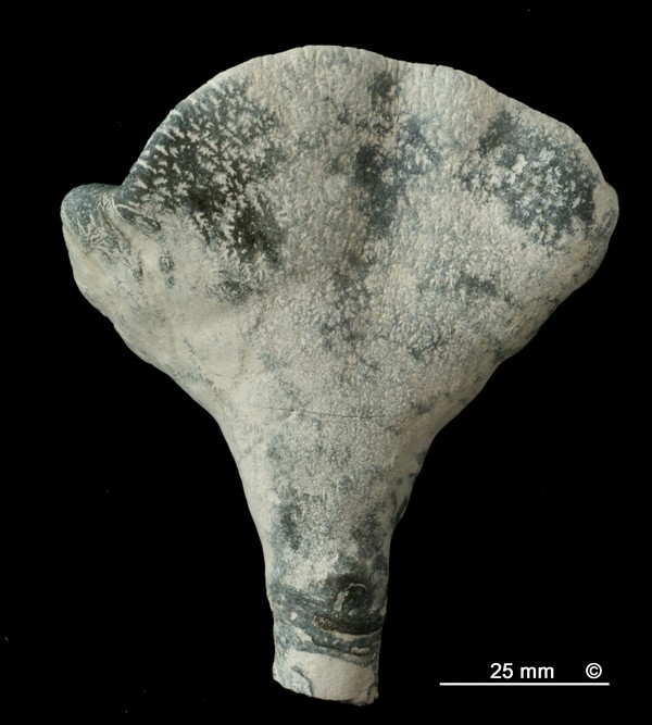
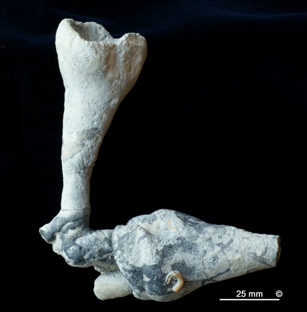
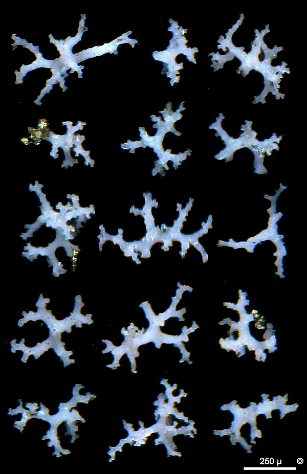
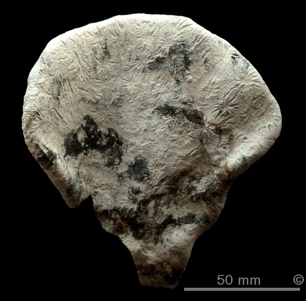
Cretaceous sponges from the Campanian of Misburg and Höver |
Figure 2 - Trachynotus auriculus, Teutonia, Misburg. Figure 3 - Trachynotus auriculus, Teutonia, Misburg. |
On the gastral surface, there are groups of small postica which are sourrounded by radiating furrows, forming large (up to 20 mm) star-like patterns (Figures 1 and 2). The pores on the outside (dermal surface) are tiny and are surrounded by mm-sized dendrite-like furrows (Figure 3). |
Figure 4 - Trachynotus auriculus, Teutonia, Misburg. |
Apparently, reproduction of Trachynotus auriculus is mainly by budding. Figure 4 shows a tilted slender funnel with three stems of junior individuals emerging from inside. The new individuals have broken off, but another one has attached itselv by formation of roots, using the senior individual as a base. |
Figure 5 - Trachynotus auriculus, Misburg. Skeleton. Figure 6 - Trachynotus auriculus, Misburg. Rhizoclones. |
The epirhyses and aporhyses of Trachynotus auriculus are up to 1 mm wide, penetrate the sponge body to about mid depth from both surfaces, and have the appearance of worm bores (See Figure 5). The skeleton of Trachynotus auriculus is very dense and strong, particularly near the dermal and gastral surfaces, and remains coherent even after acid treatment. Here, the rhizoclones are more massive and firmly joined together. Since this feature is limited to this particular species, it could due to some secondary silicification during the live-time of the sponge (i.e. cortex formation) rather than during a later diagenetic stage. The internal regions are slightly less dense (Figure 5) and single rhizoclones can be isolated by acid treatment. The skeleton consists of tiny rhizoclones (Figure 6) which tend to have isometric rather than elongate or flat shapes while curved forms are rare or absent. The rhizoclones show many side branches which connect to neighboring desmas, accounting for the high mechanical strength of the skeleton. |