
Verruculina (Amphichondrium) convoluta
Quenstedt 1878
The figure shows a neat example of Verruculina convoluta, which matches very well the descriptions given by Hinde (1883) and Schrammen (1910). The wall is approximately 5 mm thick, with a rounded margin. Small, similar sized wart-like pores cover both sides.
The species is quite rare at Misburg, but a bit more common in the Lower Campanian of Höver.
aurita_001.jpg "Locality: Alemannia, Höver.
Width: 200 mm")
Verruculina (Verruculina) aurita
Roemer 1864
The figure shows a medium-sized example of Verruculina aurita, which is a moderately common species at Misburg and Höver. Wall thickness approximately 10 mm. The inside wall surface is covered with widely spaced, approximately 1.5 mm wide postica with raised rims. Near the rounded funnel margin and on parts of the upper (paragastral) surface there are some irregularly anastomosing furrows. The outer surface shows concentric ridges (growth zones) and is covered with many, densely crowded, tiny pores, still visible by the naked eye.
aurita_004.jpg "Etched sample. Width 40 mm")
aurita_008.jpg "Etched sample. Width 40 mm")
Etched fragment of Verruculina aurita.
The upper image shows exhalent pores (postica) on the gastral surface.
The lower image shows some aporhyses leading from the postica into the interior of the sponge wall.
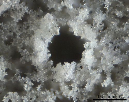
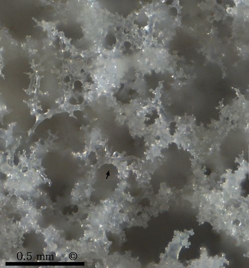
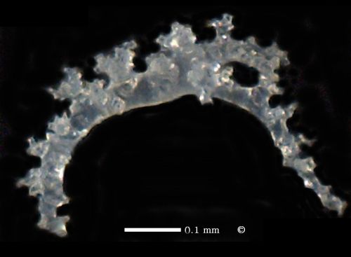
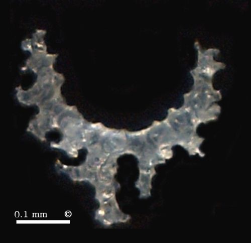
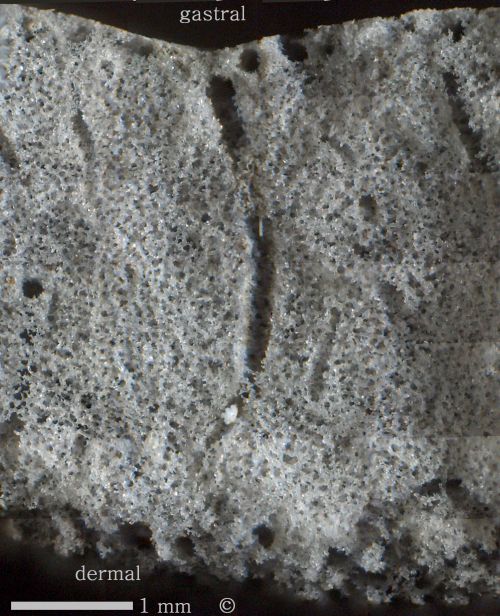
Photomicrographs of the above specimen of Verruculina aurita (etched fragment).
The upper image shows an exhalent pore with typical raised rim.
The lower image shows open canals (epirhyses) between dense skeletal parts composed of intergrown rhizoclones. Notice orientation of curved rhizoclone (arrow) with its smooth side around a canal.
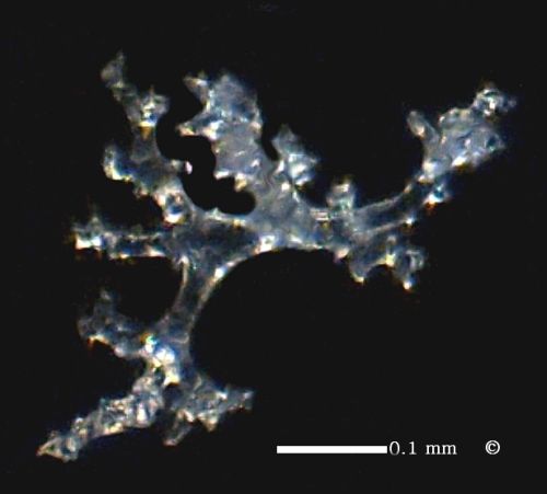
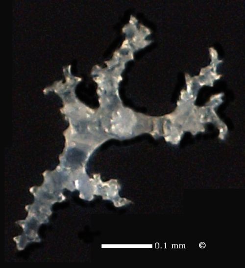
A collection of typical rhizoclone scleres of Verruculina aurita. Same etched sample as above.
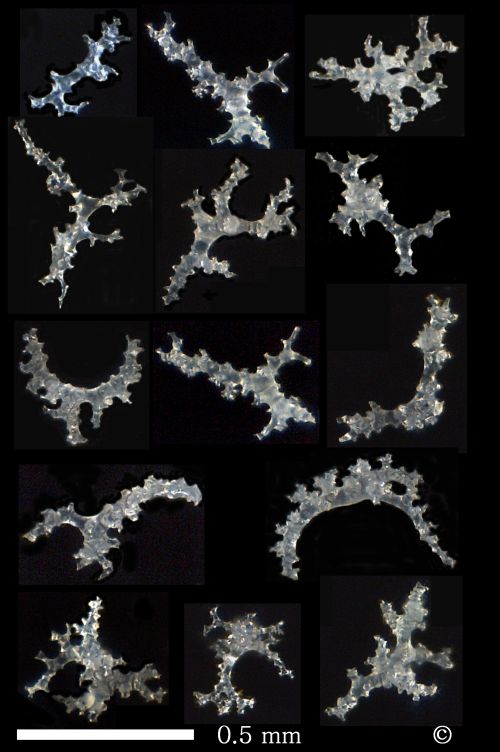
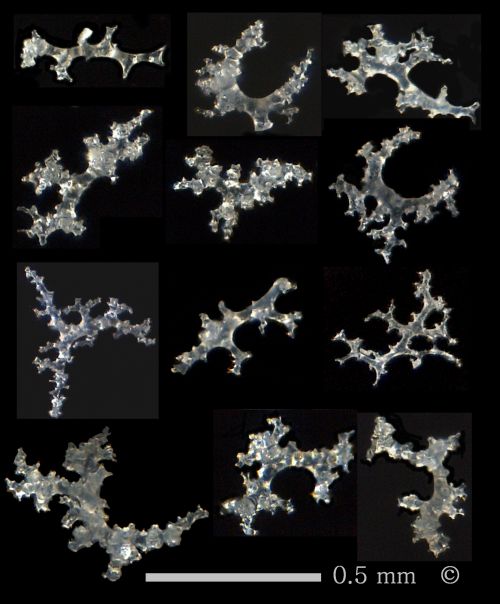
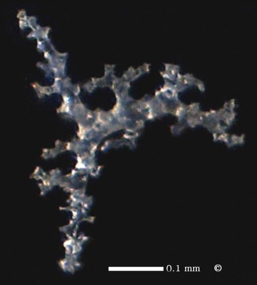
Some more rhizoclones of Verruculina aurita. Same etched sample as above.
The rhizoclones of Verruculina aurita are comparatively small. Typically, they are strongly branched in all three dimensions. Curved rhizoclones are smooth on their concave sides, and bear crescents of root-like apencices on their convex sides. The smooth, concave sides apparently correspond to channel walls, while the convex sides make the connection to the supporting skeleton.

Verruculina (Verruculina) tenuis
Roemer, 1841
Verruculina tenuis is synonymous with Chondriophyllum tenue of Schrammen (1924).
This species is moderately common at Misburg and Höver.


The ear-shaped specimen of Verruculina tenuis shown here from both sides is the most common habit to be found.
Verruculina tenuis has been characterized by Schrammen (1910) as an unpedunculated, thin-walled, ear-, plate-, or funnel-shaped sponge. (However, the author is also in posession of a specimen with a fairly long stem or straight root.)
Note the different size of the pores: The dermal side (lower image) is crowded with numerous tiny pores, barely visible by the naked eye. The gastral side (upper image) shows randomly spaced, wart-like postica, about 1 mm wide, and on average 3 to 5 mm apart. Black areas are preserved cortex.
(Serpulide worm visible on lower picture.)

Another example of Verruculina tenuis.

. . . Verruculina tenuis.
Specimens like this and the one before superficially resemble Seliscothon verrucosum. However, the wall thickness of Verruculina tenuis is less, and the radiate lamellate texture of Seliscothon is missing.

This funnel-shaped specimen is also identified as Verruculina tenuis. It has a seriate arrangement of the papillate pores along concentric lines. Its wall thickness is 5 mm at most.
_001.jpg)
_001.jpg)
Photomicrographs of of etched sample of Verruculina tenuis.
First picture: Gastral side with an exhalent pore in the center. Notice relics of cortex in the top left corner, consisting of flat siliceous platelets.
Second picture: Dermal side with several inhalent canals.
Third picture: Cross facture, showing exhalent canals (aporhyses). The curvature is convex towards the rim of the funnel.